 PREGNANCY
  GIVING BIRTH
  BABY CARE
  HEALTH & SAFETY
  FUN STUFF
  FAMILY
 Ultrasound
A pregnancy ultrasound is a non-invasive test that scans the unborn baby and the mother's reproductive organs using high frequency sound waves.
The general procedure for a pregnancy ultrasound consists of the following steps:
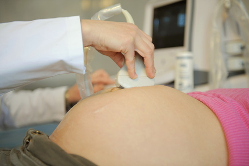
- The woman lies on a table.
- A small amount of a clear, conductive jelly is smeared on the woman's abdomen.
- The operator places the small handheld instrument called a transducer onto the woman's abdomen.
- The transducer is moved across the abdomen. The sound waves bounce off internal structures (including the baby) and are transmitted back to the transducer.)
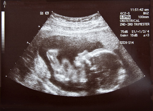
- The sound waves are then translated into a two-dimensional picture on a monitor. The mother doesn't feel or hear the transmission of the sound waves.
- By measuring the baby's body parts, such as head circumference and the length of long bones, the operator can estimate its gestational age.
This test works a bit like underwater radar, creating a picture of your baby and placenta. It is usually done in your health care provider's office, and it may require you to drink extra fluids before arriving for your appointment.
Ultrasound testing can determine the age of your baby, detect the presence of twins, help determine your due date, and measure your baby's growth and development. Sometimes an ultrasound can determine your baby's gender, but not always. Your baby's gender may or may not be clearly apparent.
Ultrasound imaging, also called ultrasound scanning or sonography, involves exposing part of the body to high-frequency sound waves to produce pictures of the inside of the body. Ultrasound exams do not use ionizing radiation (x-ray). Because ultrasound images are captured in real-time, they can show the structure and movement of the body's internal organs, as well as blood flowing through blood vessels.
Ultrasound imaging is usually a painless medical test that helps physicians diagnose and treat medical conditions.
Obstetric ultrasound provides pictures of an embryo or fetus within a woman's uterus.
A Doppler ultrasound study may be part of an obstetric ultrasound examination. Doppler ultrasound is a special ultrasound technique that evaluates blood as it flows through a blood vessel, including the body's major arteries and veins in the abdomen, arms, legs and neck.
During an obstetrical ultrasound the examiner may evaluate blood flow in the umbilical cord or may in some cases assess blood flow in the fetus or placenta.
Common Uses for Ultrasound
Apart from helping to pinpoint the unborn baby's due date, pregnancy ultrasounds are used to diagnose a number of conditions including:
- Multiple fetuses.
- Health problems with the baby.
- Ectopic pregnancy (a pregnancy outside of the uterus, most commonly, the embryo lodges in the fallopian tube instead of the uterus).
- Abnormalities of the placenta such as, placenta praevia, where the placenta is positioned over the neck of the womb (cervix).
- The health of the mother's reproductive organs.
Obstetric ultrasound should be performed only when clinically indicated to:
- establish the presence of a living embryo/fetus
- estimate the age of the pregnancy
- diagnose congenital abnormalities
- evaluate the position of the baby
- evaluate the position of the placenta
- determine if there are multiple pregnancies
- determine the amount of amniotic fluid around the baby
- check for opening or shortening of the cervix or mouth of the womb.
Ultrasound Video
Parents now commonly see ultrasound movies or images in the first trimester and clinically this is a non-invasive prenatal diagnostic tool for detection of abnormalities as well as a method of staging (ageing) and checking growth. Ultrasound can also be used in combination with other techniques to locate both embryo and placenta for other prenatal tests
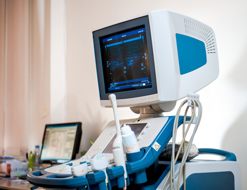
The Equipment
Ultrasound scanners consist of a console containing a computer and electronics, a video display screen and a transducer that is used to scan the body. The transducer is a small handheld device that resembles a microphone, attached to the scanner by a cord. The transducer sends out a high frequency sound wave and then listens for a returning sound wave or "echo".
The ultrasound image is immediately visible on a nearby screen that looks much like a computer or television monitor. The image is created based on the amplitude (strength), frequency and time it takes for the sound signal to return from the patient to the transducer.
Preparing for the Procedure
You should wear a loose-fitting, two-piece outfit for the examination. Only the lower abdominal area needs to be exposed during this procedure.
If an ultrasound is ordered by your clinician early in your pregnancy, you may be instructed to have a full bladder for the procedure. Air interferes with sound waves, so if your bladder is distended, the air-filled bowel is pushed out of the way by the bladder and an image of the uterus and embryo or fetus is obtained.
About an hour before the procedure you should empty your bladder. You may be instructed to drink up to six glasses of water and avoid urinating until the procedure is completed. After the first two to three months of pregnancy, a full bladder is not always necessary for imaging.
The radiologist or sonographer may elect to examine an early pregnancy by means of transvaginal ultrasound. This requires an empty urinary bladder. You should ask for specific instructions for this imaging study when you make your appointment.
How it Works
Ultrasound imaging is based on the same principles involved in the sonar used by bats, ships and fishermen. When a sound wave strikes an object, it bounces backward, or echoes. By measuring these echo waves it is possible to determine how far away the object is and its size, shape, consistency (whether the object is solid, filled with fluid, or both) and uniformity.
In medicine, ultrasound is used to detect changes in appearance and function of organs, tissues, or abnormal masses, such as tumors.
In an ultrasound examination, a transducer both sends the sound waves and records the echoing waves. When the transducer is pressed against the skin, it directs a stream of inaudible, high-frequency sound waves into the body. As the sound waves bounce off of internal organs, fluids and tissues, the sensitive microphone in the transducer records tiny changes in the sound's pitch and direction. These signature waves are instantly measured and displayed by a computer, which in turn creates a real-time picture on the monitor. These live images are usually recorded on videotape and one or more frames of the moving pictures are typically captured as still images.
The movement of the embryo or fetus and the heart beat can be seen as an ongoing ultrasound movie. Most ultrasound devices also have an audio component that processes the echoes produced by blood flowing through the fetal heart, blood vessels and umbilical cord. This sound can be made audible to human ears and has been described by patients as a whooshing noise.
Doppler ultrasound, a special application of ultrasound, measures the direction and speed of blood cells as they move through vessels. The movement of blood cells causes a change in pitch of the reflected sound waves (Doppler effect). A computer collects and processes the sounds and creates graphs or pictures that represent the flow of blood through the blood vessels.
The Procedure
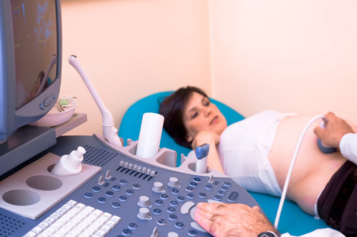
For most ultrasound exams, the patient is positioned lying face-up on an examination table that can be tilted or moved.
A clear gel is applied to the area of the body being studied to help the transducer make secure contact with the body and eliminate air pockets between the transducer and the skin. The sonographer (ultrasound technologist) or radiologist then presses the transducer firmly against the skin and sweeps it back and forth over the area of interest.
Sometimes the radiologist determines that a transvaginal scan needs to be performed. This technique often provides improved, more detailed images of the uterus and ovaries. It is especially useful in early pregnancy.
Transvaginal ultrasound is performed very much like a gynecologic exam and involves the insertion of the transducer into the vagina after the patient empties her bladder. The tip of the transducer is smaller than the standard speculum used when performing a Pap test. A protective cover is placed over the transducer, lubricated with a small amount of gel and then inserted into the vagina. Only two to three inches of the transducer end are inserted into the vagina. The images are obtained from different orientations to get the best views of the uterus and ovaries. Transvaginal ultrasound is usually performed with the patient lying on her back, possibly with her feet in stirrups similar to a gynecologic exam.
When the examination is complete, the patient may be asked to dress and wait while the ultrasound images are reviewed. However, the sonographer or radiologist is often able to review the ultrasound images in real-time as they are acquired and the patient can be released immediately.
This ultrasound examination is usually completed within 20 minutes.
Patient Experience During Ultrasound
Most ultrasound examinations are painless, fast and easy.
After you are positioned on the examination table, the radiologist or sonographer will spread some warm gel on your skin and then press the transducer firmly against your body, moving it back and forth over the area of interest until the desired images are captured. There may be varying degrees of discomfort from pressure as the transducer is pressed against the area being examined.
If scanning is performed over an area of tenderness, you may feel pressure or minor pain from the procedure.
At times the sonographer may have to press more firmly to get closer to the embryo or fetus to better visualize the structure. Any discomfort is usually minimal and temporary.
If a Doppler ultrasound study is performed, you may actually hear pulse-like sounds that change in pitch as the blood flow is monitored and measured.
With transvaginal scanning, there may be minimal discomfort as the transducer is moved in the vagina, especially when the bladder begins to refill.
Once the imaging is complete, the gel will be wiped off your skin.
After an ultrasound exam, you should be able to resume your normal activities.
Interpreting Your Ultrasound
A radiologist, a physician specifically trained to supervise and interpret radiology examinations, will analyze the images and send a signed report to your primary care or referring physician, who will share the results with you. In some cases the radiologist may discuss preliminary results with you at the conclusion of your examination.
Benefits of Ultrasound
- Ultrasound scanning is noninvasive (no needles or injections) and is usually painless.
- Ultrasound is widely available, easy-to-use and less expensive than other imaging methods.
- UItrasound imaging uses no ionizing radiation.
- Ultrasound scanning gives a clear picture of soft tissues that do not show up well on x-ray images.
- Ultrasound causes no health problems and may be repeated as often as is necessary if medically indicated.
- Ultrasound is the preferred imaging modality for the diagnosis and monitoring of pregnant women and their unborn infants.
- Ultrasound has been used to evaluate pregnancy for nearly four decades and there has been no evidence of harm to the patient, embryo or fetus. Nevertheless, ultrasound should be performed only when clinically indicated.
- Ultrasound allows the doctor to see inside the uterus and provides much information about the pregnancy.
Risks
For a standard diagnostic ultrasound there are no known harmful effects on humans.
Limitations of Ultrasound
Obstetric ultrasound cannot identify all fetal abnormalities. Consequently, when there are clinical or laboratory suspicions for a possible abnormality, a pregnant woman may have to undergo non-radiologic testing such as amniocentesis (the evaluation of fluid taken from the sac surrounding the baby) or chorionic villus sampling (evaluation of placental tissue) to determine the health of the baby, or she may be referred by her primary care provider to a perinatologist (an obstetrician specializing in high-risk pregnancies).
